Snapshot
- Tooth Preparation for Veneers: Diagnostic wax-ups, mock-ups, and smile analysis improve preparation accuracy
- Reduction Guidelines: Maintain 0.3-0.5 mm cervical and 0.5-0.7 mm mid-facial reduction for enamel-supported bonding
- Margin Accuracy: Smooth chamfer margins and rounded internal angles improve veneer fit and fabrication precision
- Functional Design: Proper incisal reduction, occlusal evaluation, and contour planning reduce fracture and debonding risks
- Digital Workflow Readiness: Clean finish lines and refined surfaces improve STL scan accuracy and CAD/CAM fabrication
- Lab Communication: Detailed scans, shade references, and preparation records help reduce remakes and adjustment issues
Synopsis
Tooth preparation for veneers should create enough space for the ceramic without unnecessarily sacrificing enamel. For most dentists, the real challenge is not aggressive reduction. It is choosing the most conservative design that still delivers predictable aesthetics, sound function, clear margins, and a restoration that the lab can actually fabricate accurately. That balance sits at the heart of long-term veneer success.
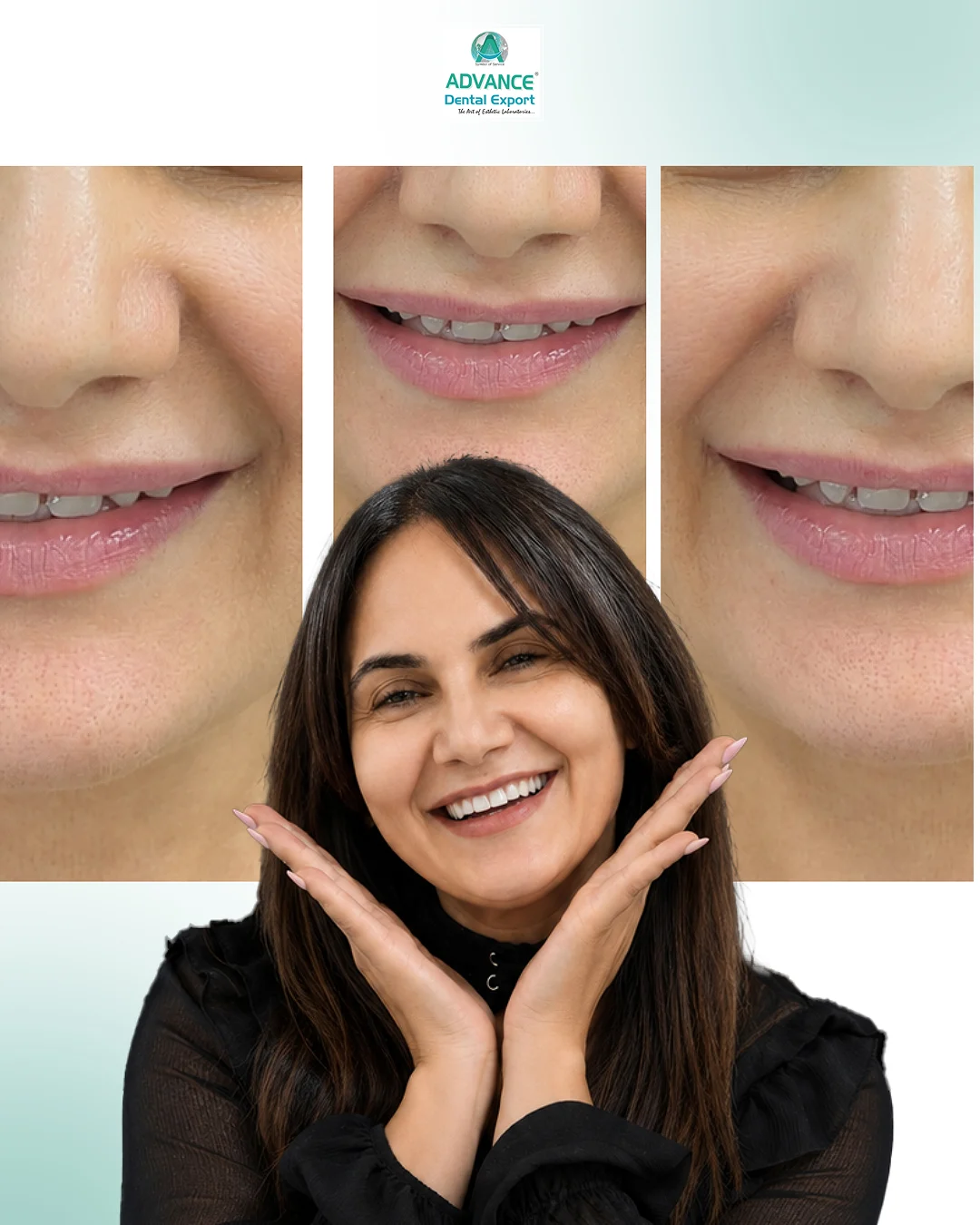
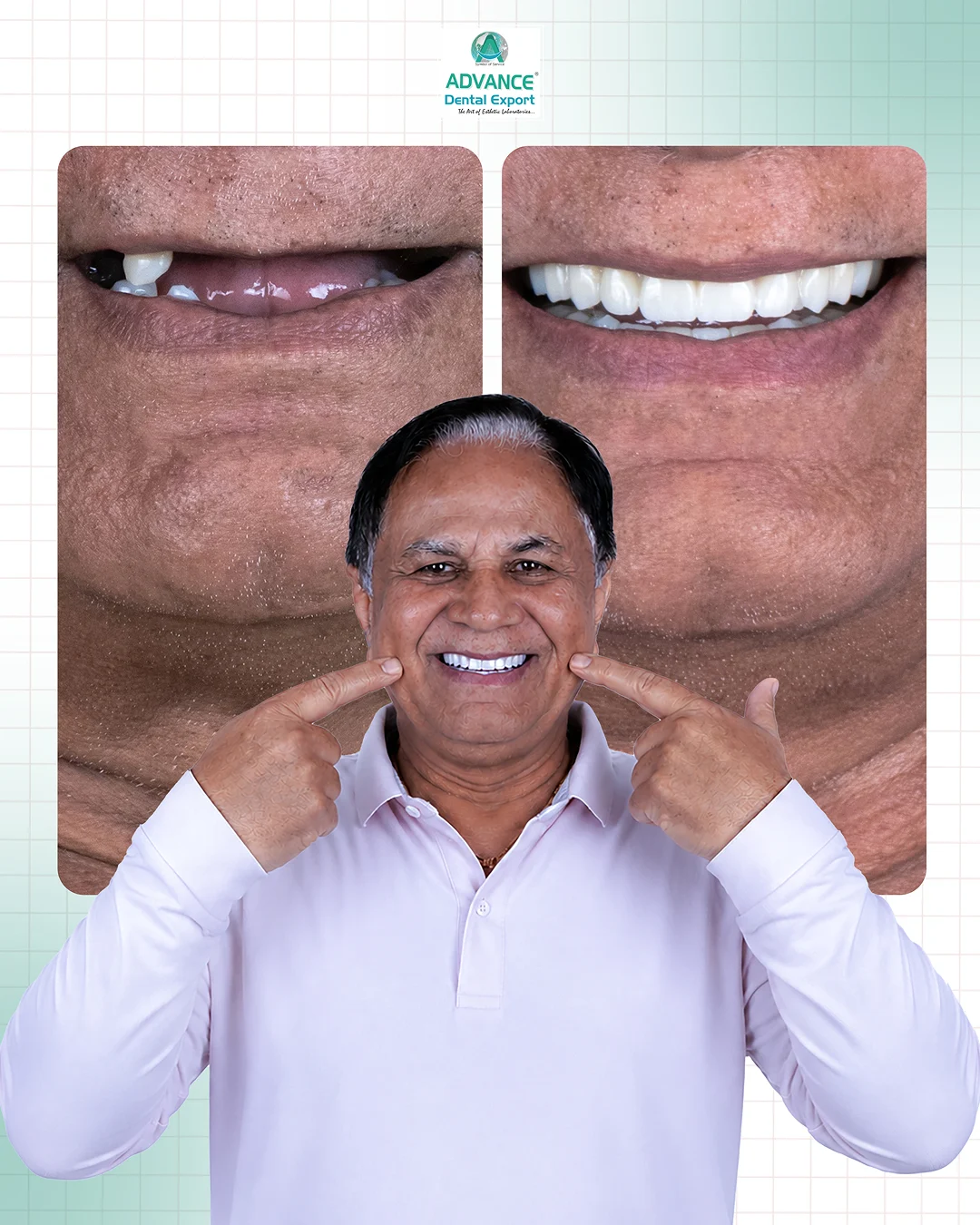
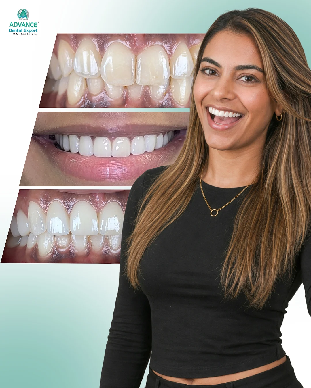
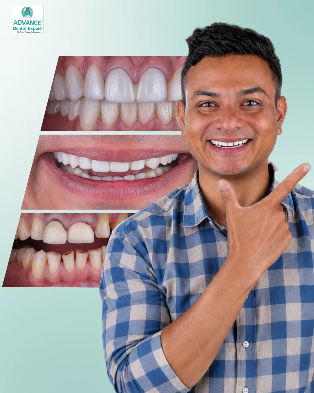
The longevity and aesthetics of veneers depend as much on preparation design as they do on ceramic selection and adhesive protocols. Even with advancements in digital dentistry , lithium disilicate materials, and CAD/CAM fabrication , veneer failures often trace back to errors made during the preparation stage.
This veneers preparation guide discusses evidence-based principles of veneer tooth preparation, with a focus on conservative approaches, preparation designs, reduction guidelines, and restorative considerations relevant to modern cosmetic dentistry.
Why Tooth Preparation Matters in Veneer Dentistry?
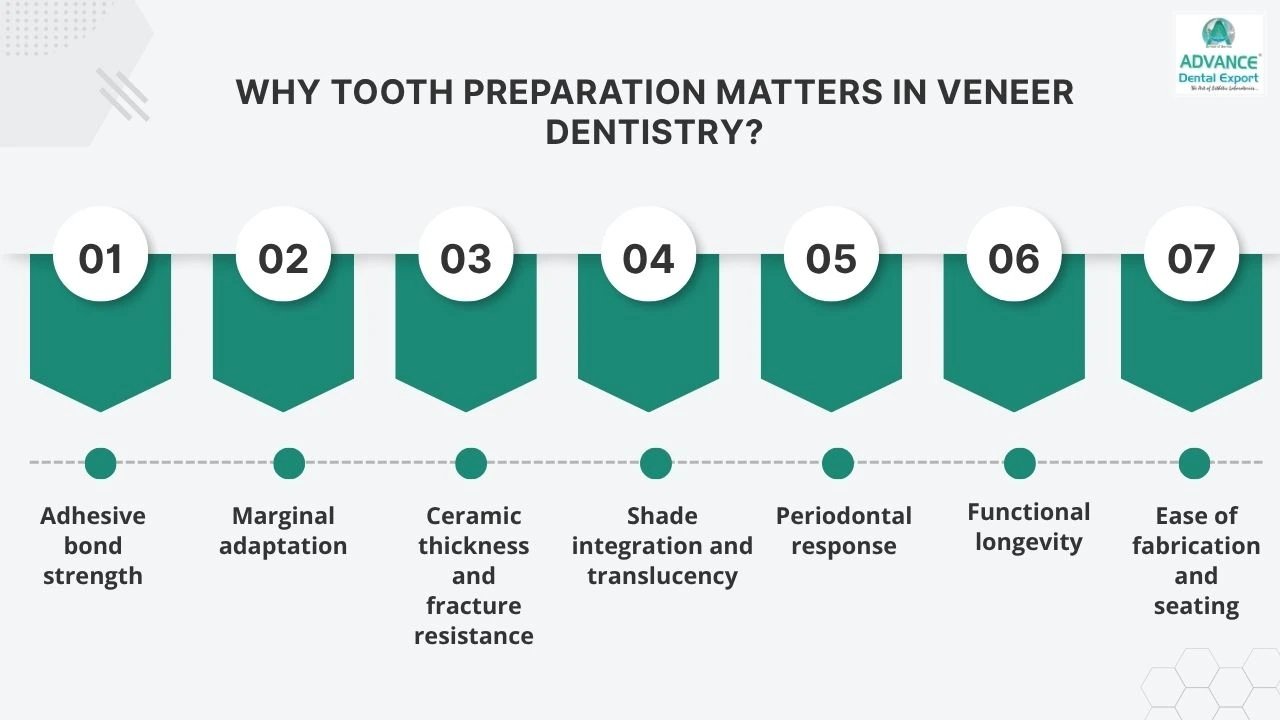
Veneer preparation influences virtually every aspect of treatment success, including:
- Adhesive bond strength
- Marginal adaptation
- Ceramic thickness and fracture resistance
- Shade integration and translucency
- Periodontal response
- Functional longevity
- Ease of fabrication and seating
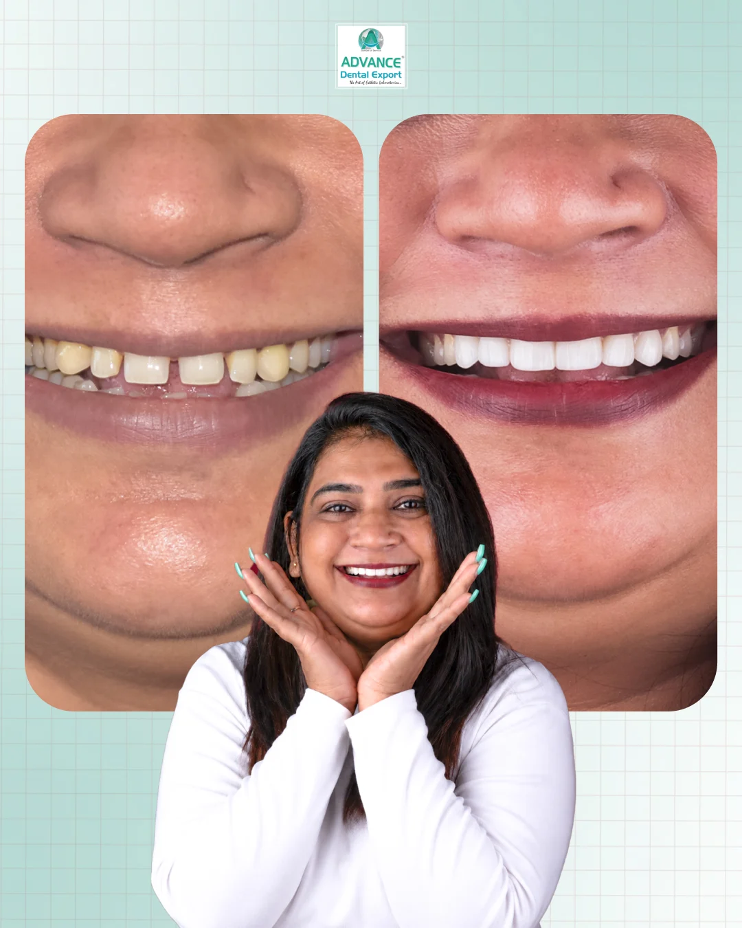
Because enamel provides the most predictable substrate for adhesive bonding, enamel preservation remains a fundamental objective during tooth preparation before veneers.
Studies consistently demonstrate improved bond durability when preparation margins remain predominantly within enamel. Consequently, clinicians should aim to preserve as much enamel as possible while achieving restorative objectives.
How Preparation Design Determines Veneer Success?
Successful veneer restorations rely heavily on tooth preparation. It controls the fit, strength, aesthetics, and durability of the restoration.
- Enamel preservation: Bond strength is highest when preparation margins remain within the enamel, improving adhesive predictability and long-term retention
- Reduction depth control: Controlled reduction creates adequate ceramic thickness without unnecessarily weakening the tooth structure
- Margin accuracy: The right type of finish line, when well-defined, has better marginal adaptation and fewer errors
- Aesthetic integration: Proper facial contouring masks defects/discolourations, improves light reflection, translucency, and natural veneer appearance
- Functional stability: Occlusal planning reduces the risk of premature contacts, fractures, and veneer debonding
Practical Tooth Reduction for Veneers
| Area | Conservative clinical guide |
| Cervical facial reduction | Around 0.3–0.5 mm in enamel-led cases |
| Mid-facial reduction | Around 0.5–0.7 mm |
| Incisal facial third | Around 0.5–0.7 mm |
| Incisal edge reduction | Often around 1.0–2.0 mm, depending on the case |
| Margin design | Light chamfer with rounded internal angles |
| Margin placement | Supra-gingival or equi-gingival, where possible |
Diagnostic Planning Before Preparing Teeth for Veneers
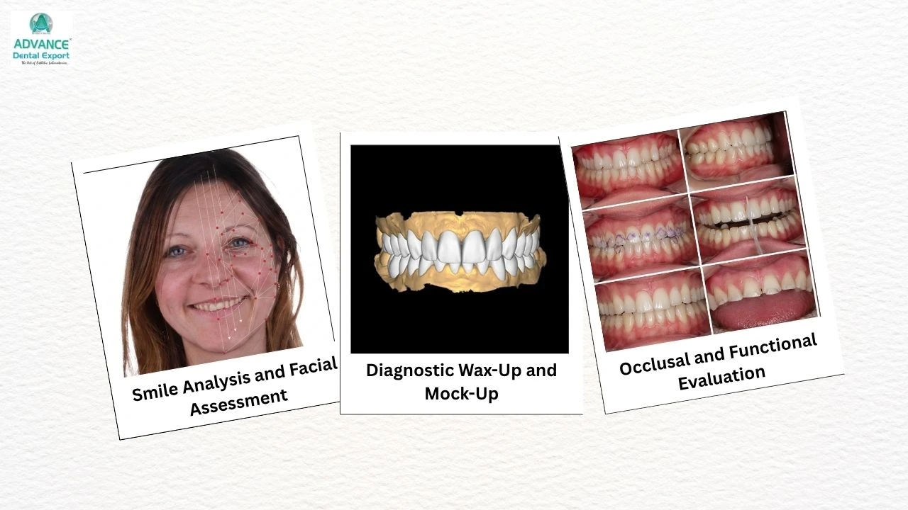
Evaluate periodontal health, tooth vitality, and existing restorations before veneer preparation, as inflamed tissue affects margin accuracy while underlying tooth condition influences preparation depth.
Understanding how are teeth prepared for veneers begins before the handpiece is activated.

Comprehensive treatment planning allows the clinician to determine whether additive, minimal-preparation, conventional preparation, or even alternative restorative approaches are most appropriate.
Smile Analysis and Facial Assessment
Preparation design should support the desired aesthetic outcome rather than dictate it.
Clinical assessment should include:
- Smile line evaluation
- Gingival symmetry
- Tooth proportions
- Midline analysis
- Buccal corridor assessment
- Incisal edge position
- Lip mobility during speech and smiling
- Facial harmony and profile considerations
These observations help establish restorative goals and determine the amount of reduction required.
Diagnostic Wax-Up and Mock-Up
Diagnostic wax-ups and mock-ups serve as reference points for facial and dental proportions and reduction depth. This helps to visualise the final smile design before preparation begins.
They help clinicians:
- Visualize the final restorative outcome
- Evaluate aesthetics and phonetics
- Assess incisal position
- Communicate treatment expectations
- Guide conservative enamel preparation
- Identify areas requiring selective reduction
Occlusal and Functional Evaluation
Functional analysis should never be overlooked during veneer treatment planning.
- Static occlusion
- Anterior guidance
- Excursive movements
- Parafunctional habits
- Existing wear patterns
- Edge-to-edge relationships
Tooth Reduction for Veneers: Recommended Guidelines
Controlled tooth reduction for veneers allows adequate ceramic thickness without sacrificing unnecessary tooth structure.
Preparation requirements vary depending on material choice , tooth colour, and restorative objectives.
Facial Reduction
Facial reduction should follow natural tooth anatomy. A two-plane approach is generally preferred. Recommended reduction guidelines include:
- Cervical third: approximately 0.3–0.5 mm
- Middle facial region: approximately 0.5–0.7 mm
- Incisal facial third: approximately 0.5–0.7 mm
Benefits of two-plane facial reduction include:
- Better preservation of natural contours
- Uniform ceramic thickness
- Improved shade integration
- Reduced risk of over-contouring
Depth-cutting burs and silicone indices assist in maintaining accuracy throughout preparation.
Incisal Reduction Design
Incisal preparation remains one of the most debated aspects of dental preparation for veneers. The selected design should consider aesthetics, function, insertion path, and material requirements.
Recommended incisal reduction typically ranges from 1.0–2.0 mm, depending on:
- Material selection
- Desired translucency
- Occlusal considerations
- Incisal edge modifications
Margin Design and Finish Line Accuracy
Margin quality directly affects seating accuracy and marginal adaptation during veneer fabrication.
- Smooth chamfer margins improve adaptation, seating, and fabrication precision
- Feather-edge margins increase the risk of over-contoured restorations and marginal discrepancies
- Poorly placed margins can affect aesthetics, hygiene maintenance, and periodontal response
How to Achieve:
- Maintain smooth chamfer finish lines with rounded internal angles
- Evaluate finish lines from the incisal or occlusal view before final impression or scanning
- Place margins supra-gingivally or equi-gingivally whenever clinically possible
Interproximal Preparation Control
Interproximal preparation significantly influences emergence profile and aesthetic outcomes. The decision to preserve or break contact depends on treatment objectives.
Preserve Contacts When:
- Alignment is acceptable
- Emergence profiles require minimal modification
- Diastema closure is unnecessary
- Maximum enamel conservation is desired
Break Contacts When:
- Correcting tooth shape
- Closing spaces
- Managing discolouration extending interproximally
- Altering the emergence profile
- Addressing alignment discrepancies
Excessive interproximal reduction should be avoided because it unnecessarily sacrifices enamel. Fine diamond burs and abrasive strips provide controlled access during conservative preparation.
Finish Line Design and Margin Placement
Margin quality directly affects adaptation, periodontal health, and digital accuracy.
Chamfer Margin Design
A light chamfer margin is often preferred.
Advantages include:
- Clear margin identification
- Improved ceramic adaptation
- Predictable laboratory fabrication
- Reduced stress concentration
Margins should demonstrate:
- Rounded internal angles
- Smooth transitions
- Consistent depth
Avoid Feather-Edge Margins
Feather-edge designs may contribute to:
- Over-contoured restorations
- Margin distortion
- Fabrication difficulties
- Reduced seating accuracy
Margin Placement
Whenever clinically appropriate, margins should remain:
- Supra-gingival, or
- Equi-gingival.
Subgingival extension may be indicated when:
- Masking cervical discoloration
- Managing existing restorations
- Addressing aesthetic concerns involving visible margins
However, unnecessary subgingival preparation increases challenges related to moisture control and periodontal health.
Finish Line Design and Margin Placement
Preparation finishing influences both impression accuracy and veneer adaptation.
- Rough surfaces and unsupported enamel compromise marginal adaptation
- Sharp line angles increase stress concentration within ceramic restorations
- Surface irregularities can reduce digital scan accuracy and impression precision
How to Achieve:
- Smooth preparation surfaces using fine finishing burs before impression or scanning
- Round internal line angles to improve ceramic adaptation and stress distribution
Digital Workflow Considerations for Predictable Veneers
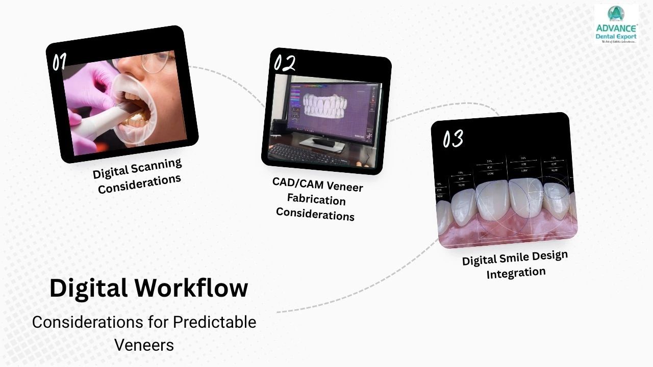
Digital workflows improve precision only when tooth preparation quality supports accurate scanning and fabrication. Poor finish lines or inconsistent reduction can still compromise the final restoration.
Digital Scanning Considerations
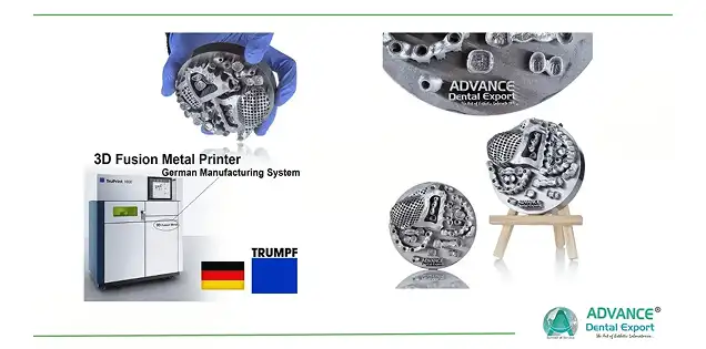
Looking for a trusted lab partner? Get high-precision restorations from Advance Dental Export
Digital scans depend heavily on preparation and margin clarity.
- Clearly defined margins
- Smooth preparation surfaces
- Effective moisture control
- Proper tissue management
- Adequate access
Poor finish lines compromise STL capture and subsequent restoration design.
CAD/CAM Veneer Fabrication Considerations
Preparation design directly affects milling accuracy and ceramic strength in CAD/CAM workflows . Preparation design influences:
- Milling accuracy
- Internal adaptation
- Ceramic thickness consistency
- Shade predictability
- Restoration strength
Digital Smile Design Integration
Digital smile design improves visualisation and communication throughout the veneer planning process.
- Helps align preparation design with the planned restorative outcome
- Improves communication between the clinician, patient, and lab
- Allows better evaluation of contour, incisal position, and smile balance before fabrication
Role of Dental Laboratory in Predictable Veneer Fabrication
Successful veneer cases rely on collaboration between the clinician and the dental laboratory. Providing detailed information reduces remakes and improves predictability. Essential records include:
- STL files or impressions
- Preparation photographs
- Diagnostic wax-up references
- Mock-up images
- Shade documentation
- Surface texture references
- Occlusal records
- Smile design instructions
Common Preparation Errors to Avoid
- Excessive facial reduction
- Unnecessary dentin exposure
- Inadequate reduction resulting in bulky veneers
- Poorly defined finish lines
- Sharp internal angles
- Ignoring occlusal considerations
- Overextending interproximal preparations
- Failing to use reduction guides
- Inadequate communication with the laboratory
Take Home Message
Predictable tooth preparation for veneers requires far more than achieving a target reduction depth. Every decision—from diagnostic planning and smile analysis to margin placement and finishing—affects the final restoration's aesthetics, function, and longevity.
Conservative preparation principles centred on enamel preservation continue to provide the most reliable foundation for adhesive success. Controlled tooth reduction, appropriate finish line design, thoughtful incisal preparation, and careful occlusal assessment help clinicians balance biological preservation with restorative demands.
Whether fabricating conventional porcelain veneers or lithium disilicate restorations such as Emax veneers, meticulous preparation remains one of the most influential determinants of long-term clinical outcomes in cosmetic dentistry .
Answers in Brief for Your Queries
How are teeth prepared for veneers?
Teeth are prepared using conservative reduction principles that preserve enamel while creating sufficient space for ceramic. Preparation typically includes facial reduction, incisal design selection, margin refinement, and evaluation of occlusion and aesthetics.
How much tooth reduction is recommended for veneers?
Facial reduction generally ranges from 0.3–0.5 mm cervically and 0.5–0.7 mm in the mid-facial region. Incisal reduction often ranges from 1.0 to 2.0 mm, depending on restorative goals and material selection.
Why is enamel preservation important during veneer preparation?
Bonding to enamel provides superior adhesion, improved marginal integrity, greater long-term predictability, and reduced postoperative sensitivity compared with extensive dentin exposure.
What is the preferred finish line design for veneers?
A light chamfer margin with rounded internal line angles is commonly recommended because it improves marginal adaptation, scanning accuracy, and laboratory fabrication precision.
When should interproximal contacts be broken during veneer preparation?
Contacts may be broken when correcting contour discrepancies, closing diastemas, masking interproximal discoloration, or modifying emergence profiles. Otherwise, preserving contacts supports enamel conservation.
Can no-prep veneers be used in all cases?
No. No-prep veneers are suitable only in carefully selected cases involving favourable tooth position and minimal restorative changes. They are not appropriate for every cosmetic indication.